Body
Human islets display unique morphological and functional differences compared to rodent models; therefore, they are a critical resource for better understanding diabetes physiology in humans. Due to limited supply and increased researcher needs, LifeNet Health LifeSciences has begun developing protocols to isolate high-quality human islets.
![Isolated human islets stained with fluorescein diacetate (FDA) and propidium iodide (PI)](https://cdn.lnhlifesciences.org/sites/default/files/2020-11/Islet%20viability_FDA%20PI_new.jpg)
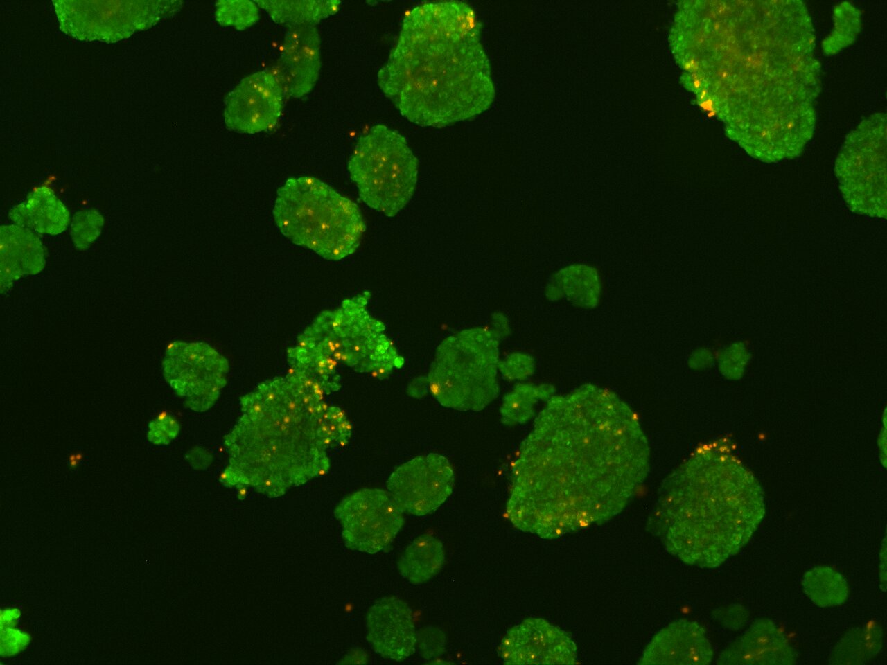
Islets shown with viability stains FDA and PI
![Isolated human islet stained for insulin and glucagon](https://cdn.lnhlifesciences.org/sites/default/files/2020-08/ICC_Islet_Insulin%28G%29-Glucagon%28R%29_20x-1.jpg)
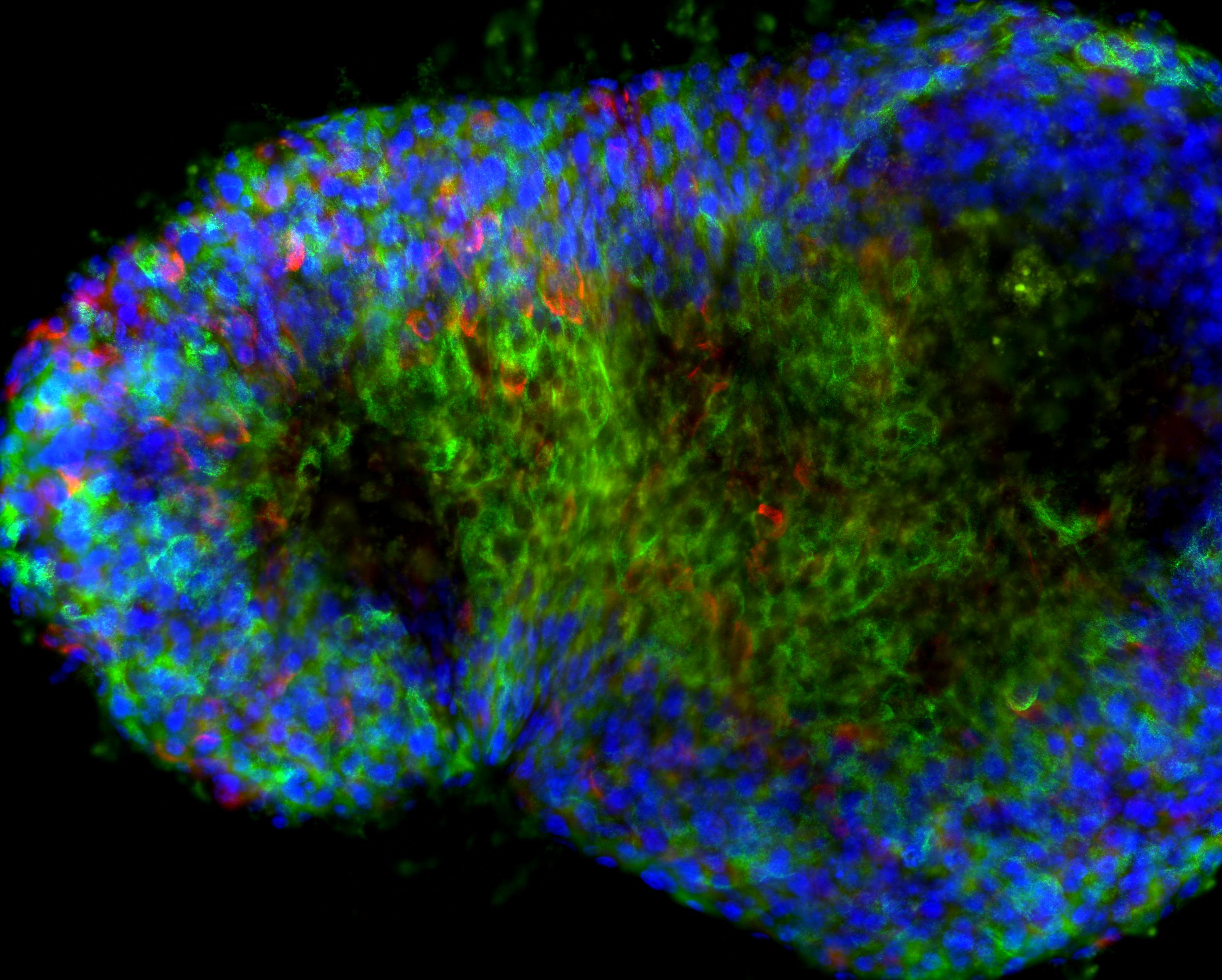
Islets stained for Insulin and Glucagon
![Isolated human islets on Day 12 of culture stained with diphenylthiocarbazone (DZT)](https://cdn.lnhlifesciences.org/sites/default/files/2020-08/Islets_D12%20post-culture-1.jpg)
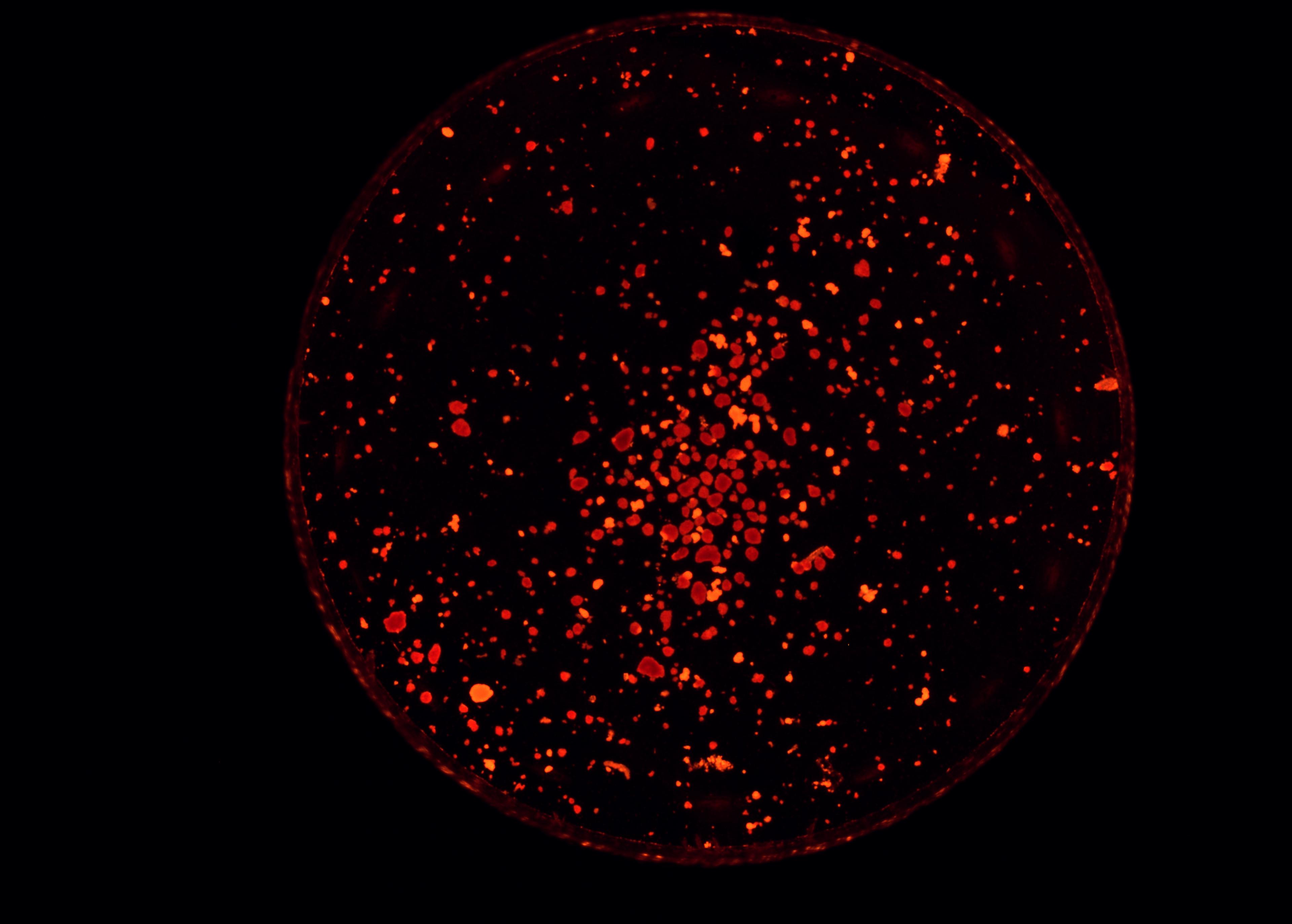
Islets in Culture stained with diphenylthiocarbazone-Day 12
![H&E stained image of human pancreatic tissue showing intact islet](https://cdn.lnhlifesciences.org/sites/default/files/2020-08/Histology_Pancreas_Normal_10x-2.jpg)
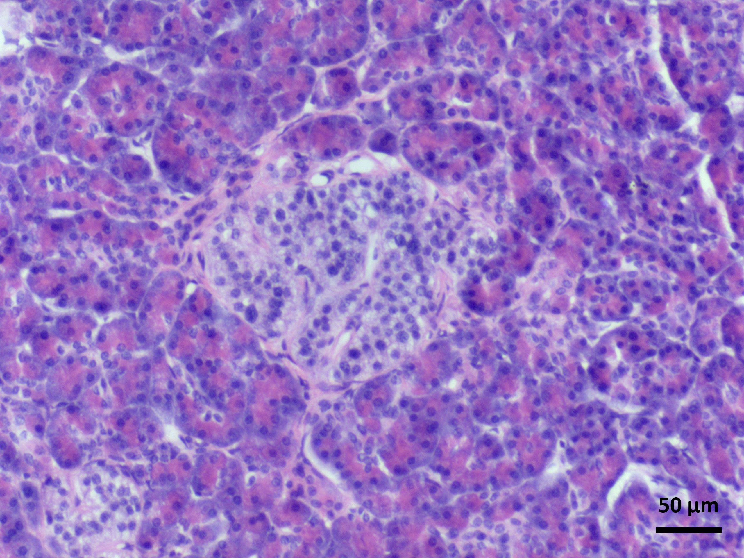
H&E Stained Pancreatic Islet
![Immunohistochemical (IHC) stained human pancreatic tissue with islet stained for the secretion of insulin and amylase](https://cdn.lnhlifesciences.org/sites/default/files/2020-08/IHC_Pancreas_Insulin%28G%29-Amylase%28R%29_20x.jpg)
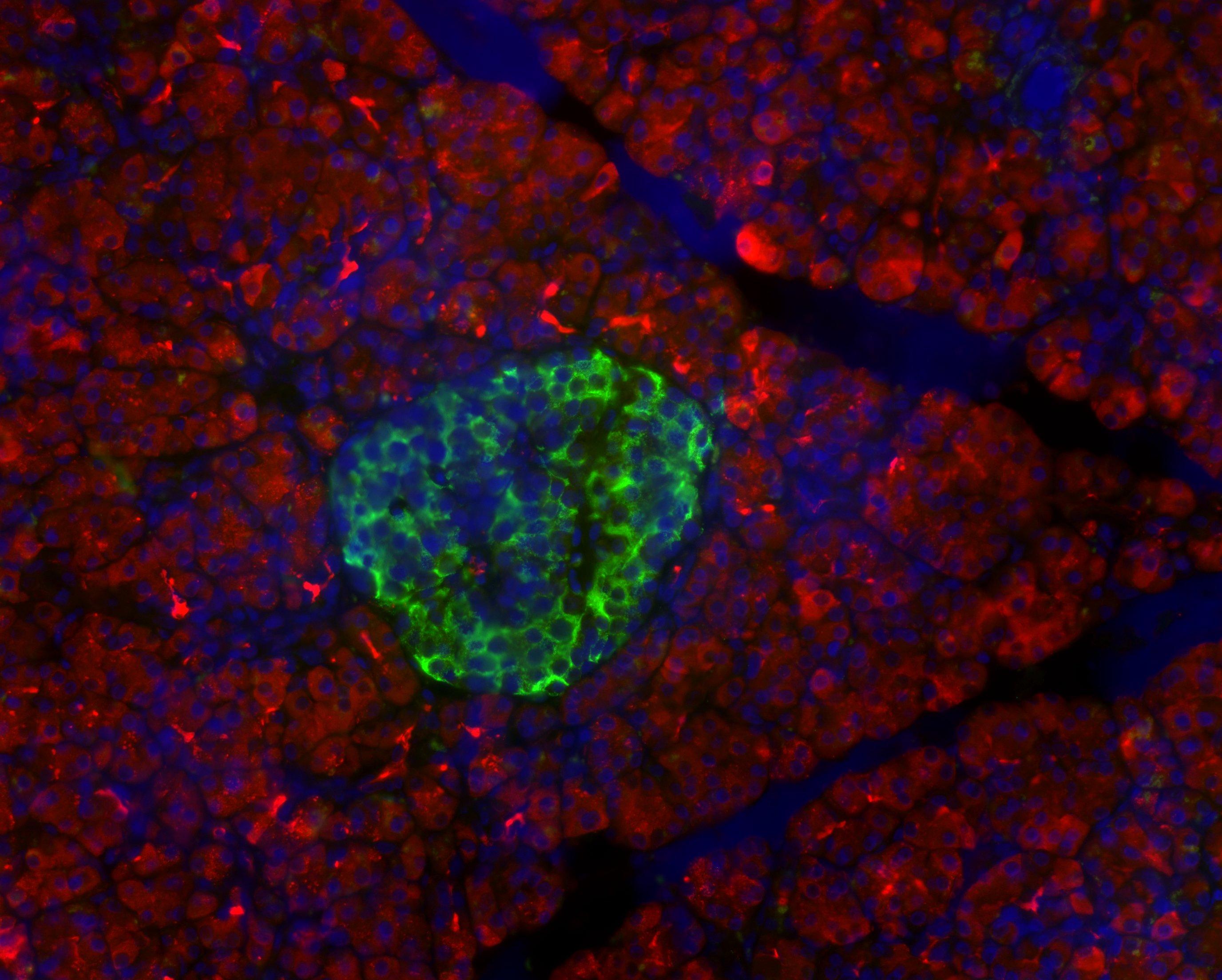
Pancreatic Islet cell grouping stained with antibodies for insulin and amylase
![Immunohistochemical (IHC) stained human pancreatic tissue with islet stained for the secretion of insulin and glucagon](https://cdn.lnhlifesciences.org/sites/default/files/2020-08/IHC_Insulin%28G%29-Glucagon%28R%29_10x.jpeg)
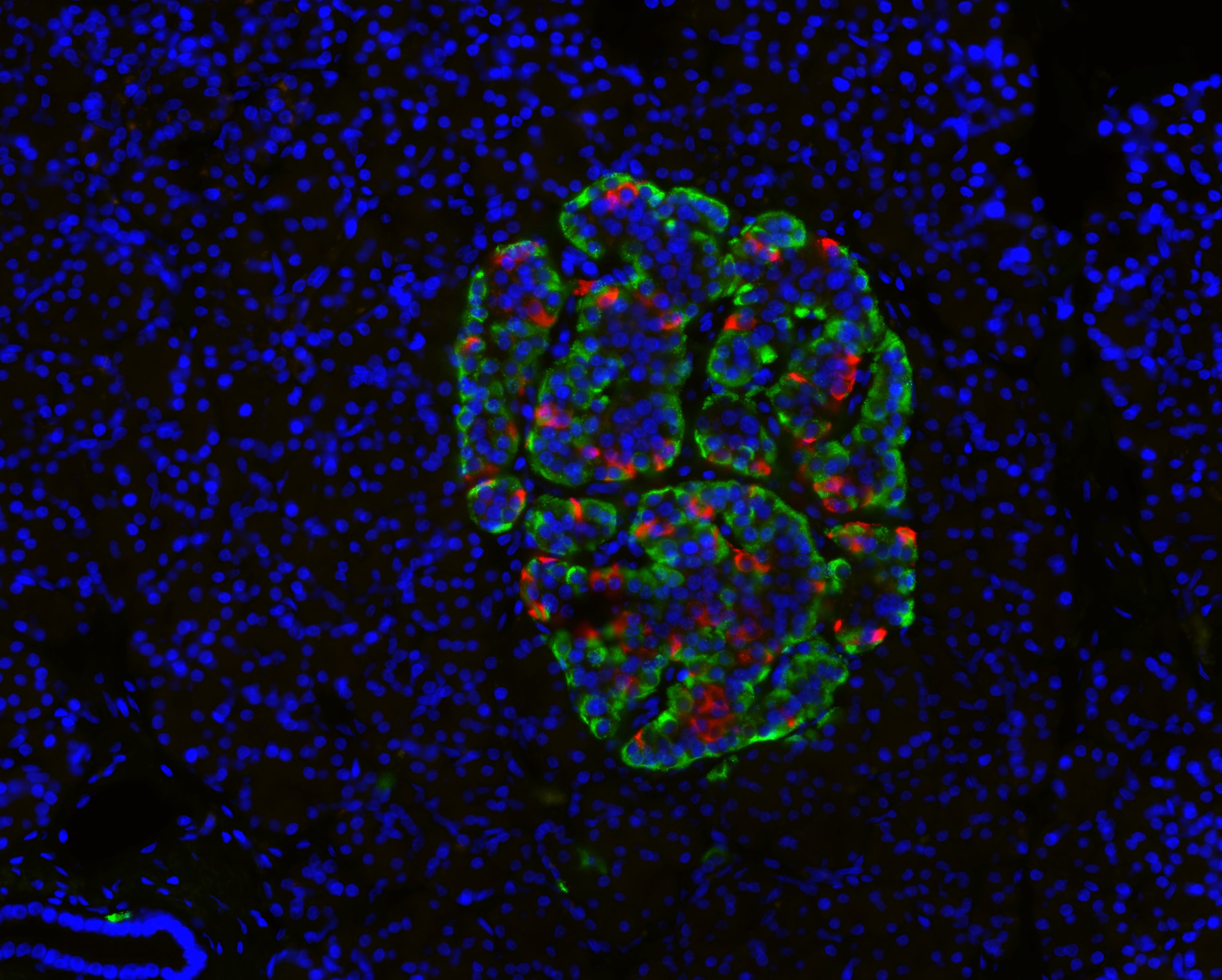
Pancreatic Islet cell grouping stained with antibodies for insulin and glucagon