Cytotoxicity Assays Can Improve Compound Safety
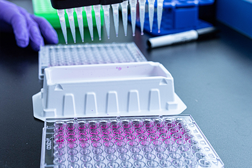
Determining cytotoxicity, or harm to cells, is a critical component of identifying the viability of new chemical entities (NCEs) and other compounds early in the preclinical phase of drug development or product discovery. Cytotoxicity assays measure cell viability, offering researchers key insight into potential adverse effects.
This is an important step that can help save time and money. The drug and chemical development process is expensive and drawn out. Drug development, for example, can take up to 12 years. Failure of new drug candidates due to unforeseen toxicity wastes valuable time and resources.
Agrichemical development can also be a complex process. Understanding potential cytotoxic properties – including endocrine disruption – at relevant exposure concentrations can also help inform Next Generation Risk Assessments (NGRAs).
Whatever the industry, in vitro cytotoxicity assays can greatly improve the success of new compounds in animal safety studies, human clinical trials, and NGRAs.
LifeNet Health LifeSciences offers carefully designed in vitro cytotoxicity tests and screening models that are mechanism-based. As a result, we can provide information on the relative toxicity of individual NCEs – with higher-throughput options to assess full compound libraries. We can work with your team to monitor changes in toxicity as you optimize the candidate molecule.
We support:
- Drug discovery, with special expertise in small molecules, nanoparticles, antibody drug conjugates (ADCs), oligonucleotides, and antisense oligonucleotides (ASOs). Our assays can reduce the risk of late-stage attrition due to safety.
- Agrichemical development.
- Cosmetics and personal care safety.
- Food additive safety.
We are committed to high scientific quality and timely turnaround.
Connect with an expert
Tests We Offer
Assays
- Multiple End Point Toxicity Testing (endpoint panels custom designed for our clients)
- Membrane integrity
- MitoTox – Gluc/Gal, ATP, MTT, mitochondrial membrane potential
- Oxidative stress
- Inflammatory response
- Apoptosis/pyroptosis
Test Models (2D and 3D organotypic)
- Intestine
- Liver
- Kidney
- Lung
- Multiple primary cells and cell lines
- TruVivo®
Harness the TruVivo system
Cytotoxicity assays can be performed using TruVivo, our innovative hepatic system that provides human-relevant, reliable results. Advantages include:
- Greater human relevance thanks to TruVivo's ability to mimic the microarchitecture and basic functionality of the human liver. The formation of hepatocyte colonies, with cell-to-cell interactions and junctional complexes, are key advantages compared to standard monocultures.
- Longer-term studies (>14 days) compared to sandwich cultures or suspension assays. This is a helpful option to consider for compounds, where lower doses repeated exposures for extended periods are required.
- Greater reliability, with accurate, consistent results. TruVivo’s reliability is proven by consistent urea and albumin production from well to well, experiment to experiment, and user to user.
Our Process Sets Us Apart From Other Laboratories
Consultation – We partner with you to determine the best approach, offering study plans that draw from our 60-plus years of combined in vitro testing experience. We may recommend utilizing more than one assay when performing a thorough cytotoxicity screening.
Assay Development – We develop and validate your assay using many different cell models, with an emphasis on all-human primary cells and biospecimens to simulate in vitro conditions, which provides more relevant, reliable results than traditional testing methods. This is part of our commitment to advancing alternative methods to support organizations in moving away from traditional animal-based models.
Data Analysis – We provide expert data analysis, including offering adaptable options when needed based on early results.
Results Review – Our clear, concise reports bring simplicity to your biggest data challenges, putting your organization in the best position for regulatory reviews. We may suggest follow-up studies to further your organization’s research goals.
We offer extensive testing capabilities. Our facilities are compliant with Good Laboratory Practices and Good In Vitro Method Practices (OECD 286).